Search the site...

Amada Mona
  • Blog
  • About
  • Contact
  • Blog
  • About
  • Contact

What Is Pes Planus?

7/3/2017

 
Overview

Acquired Flat Feet

The condition known as ?fallen arches? is also referred to as flat feet or pes planus. It arises when the arch of the foot collapses to the point that the entire sole is flat to the ground. It is commonly seen in young children who have yet to develop the muscles in the sole of their feet. In most cases as a child develops and learns how to walk the finer intrinsic muscles that support the sole of the foot will develop normally and an arch forms. However in a small number of cases these muscles don?t form properly and thus neither does the arch. Fallen arches which are acquired in adulthood may be due to a number of factors.

Causes

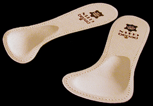
Just as there are many different causes of flat feet, there are also many different treatment options. The most important aspect of treatment is determining the exact type or underlying cause of flat feet that you have. Foot and ankle specialists can determine this through thorough clinical examination and special imaging studies (e.g., x-rays, computed tomography, and/or magnetic resonance imaging). Conservative treatment is effective in the vast majority of flat foot cases, and consists of things such as insoles, splints, manipulation, or casting. Surgery is required much less frequently, and is reserved only for some of the severe types of flat foot that do not respond to conservative therapy. You may have noticed that one common element in the conservative treatment of all types of flat feet is orthoses. Many companies now manufacture semi-custom orthotic devices that not only improve comfort, but also seek to control abnormal motion of the foot. These over-the-counter inserts, in the $25 to $50 range, are an economical treatment that may help a majority of people. Unfortunately, these semi-custom devices will not fit everyone perfectly, and those of us who differ too much from the average may respond better to custom orthotic devices. Custom inserts are prescribed by your foot and ankle specialist and are made individually from either a physical or computerized impression of your feet. The only drawback of custom orthoses is their cost, ranging anywhere from $300 to $500. Many physicians recommend trying over-the-counter inserts first (and even keep them in stock) as they may save their patients large sums of money.

Symptoms

Depending on the cause of the flatfoot, a patient may experience one or more of the different symptoms below. Pain along the course of the posterior tibial tendon which lies on the inside of the foot and ankle. This can be associated with swelling on the inside of the ankle. Pain that is worse with activity. High intensity or impact activities, such as running, can be very difficult. Some patients can have difficulty walking or even standing for long periods of time. When the foot collapses, the heel bone may shift position and put pressure on the outside ankle bone (fibula). This can cause pain on the outside of the ankle. Arthritis in the heel also causes this same type of pain. Patients with an old injury or arthritis in the middle of the foot can have painful, bony bumps on the top and inside of the foot. These make shoewear very difficult. Occasionally, the bony spurs are so large that they pinch the nerves which can result in numbness and tingling on the top of the foot and into the toes. Diabetics may only notice swelling or a large bump on the bottom of the foot. Because their sensation is affected, people with diabetes may not have any pain. The large bump can cause skin problems and an ulcer (a sore that does not heal) may develop if proper diabetic shoewear is not used.

Diagnosis

An examination of the foot is enough for the health care provider to diagnose flat foot. However, the cause must be determined. If an arch develops when the patient stands on his or her toes, the flat foot is called flexible and no treatment or further work-up is necessary. If there is pain associated with the foot or if the arch does not develop with toe-standing, x-rays are necessary. If a tarsal coalition is suspected, a CT scan is often ordered. If a posterior tibial tendon injury is suspected, your health care provider may recommend an MRI.

bestshoelifts

Non Surgical Treatment

Normally, flat feet disappear by age six as the feet become less flexible and the arches develop. Only about 1 or 2 out of every 10 children will continue to have flat feet into adulthood. For children who do not develop an arch, treatment is not recommended unless the foot is stiff or painful. Shoe inserts won?t help your child develop an arch, and may cause more problems than the flat feet themselves. However, certain forms of flat feet may need to be treated differently. For instance, a child may have tightness of the heel cord (Achilles tendon) that limits the motion of his foot. This tightness can result in a flat foot, but it usually can be treated with special stretching exercises to lengthen the heel cord. Rarely, a child will have truly rigid flat feet, a condition that can cause problems.

Surgical Treatment

Adult Acquired Flat Foot

A better approach is to strengthen the weakened ligaments with Prolotherapy, supplemented by an arch support if the condition has existed for several years. Chronic pain is most commonly due to tendon and ligament weakness, or cartilage deterioration. The safest and most effective natural medicine treatment for repairing tendon, ligament and cartilage damage is Prolotherapy. In simple terms, Prolotherapy stimulates the body to repair painful areas. It does so by inducing a mild inflammatory reaction in the weakened ligaments and cartilage. Since the body heals by inflammation, Prolotherapy stimulates healing. Prolotherapy offers the most curative results in treating chronic pain. It effectively eliminates pain because it attacks the source: the fibro-osseous junction, an area rich in sensory nerves. What?s more, the tissue strengthening and pain relief stimulated by Prolotherapy is permanent.

After Care

Patients may go home the day of surgery or they may require an overnight hospital stay. The leg will be placed in a splint or cast and should be kept elevated for the first two weeks. At that point, sutures are removed. A new cast or a removable boot is then placed. It is important that patients do not put any weight on the corrected foot for six to eight weeks following the operation. Patients may begin bearing weight at eight weeks and usually progress to full weightbearing by 10 to 12 weeks. For some patients, weightbearing requires additional time. After 12 weeks, patients commonly can transition to wearing a shoe. Inserts and ankle braces are often used. Physical therapy may be recommended. There are complications that relate to surgery in general. These include the risks associated with anesthesia, infection, damage to nerves and blood vessels, and bleeding or blood clots. Complications following flatfoot surgery may include wound breakdown or nonunion (incomplete healing of the bones). These complications often can be prevented with proper wound care and rehabilitation. Occasionally, patients may notice some discomfort due to prominent hardware. Removal of hardware can be done at a later time if this is an issue. The overall complication rates for flatfoot surgery are low.

Leg Length Discrepancy Following Knee Replacement Surgery

6/30/2017

 
Overview

Many people don?t realise it, but one of their legs is longer (or shorter) than the other one. Over time, this can lead to degenerative osteoarthritis (OA) in the hip joint requiring a hip replacement. But the surgeon can?t just take the old hip joint out and put the new implant in. Careful planning, special surgical techniques, and adjusting of the implant component parts are important in preventing continuation or even worsening of the leg length discrepancy.Leg Length Discrepancy

Causes

The causes of LLD are many, including a previous injury, bone infection, bone diseases (dysplasias), inflammation (arthritis) and neurologic conditions. Previously broken bones may cause LLD by healing in a shortened position, especially if the bone was broken in many pieces (comminuted) or if skin and muscle tissue around the bone were severely injured and exposed (open fracture). Broken bones in children sometimes grow faster for several years after healing, causing the injured bone to become longer. Also, a break in a child?s bone through a growth center (located near the ends of the bone) may cause slower growth, resulting in a shorter extremity. Bone infections that occur in children while they are growing may cause a significant LLD, especially during infancy. Bone diseases may cause LLD, as well; examples are neurofibromatosis, multiple hereditary exostoses and Ollier disease. Inflammation of joints during growth may cause unequal extremity length. One example is juvenile rheumatoid arthritis. Osteoarthritis, the joint degeneration that occurs in adults, very rarely causes a significant LLD.

Symptoms

The patient/athlete may present with an altered gait (such as limping) and/or scoliosis and/or low back pain. Lower extremity disorders are possibly associated with LLD, some of these are increased hip pain and degeneration (especially involving the long leg). Increased risk of: knee injury, ITB syndrome, pronation and plantar fascitis, asymmetrical strength in lower extremity. Increased disc or vertebral degeneration. Symptoms vary between patients, some patients may complain of just headaches.

Diagnosis

The most accurate method to identify leg (limb) length inequality (discrepancy) is through radiography. It?s also the best way to differentiate an anatomical from a functional limb length inequality. Radiography, A single exposure of the standing subject, imaging the entire lower extremity. Limitations are an inherent inaccuracy in patients with hip or knee flexion contracture and the technique is subject to a magnification error. Computed Tomography (CT-scan), It has no greater accuracy compared to the standard radiography. The increased cost for CT-scan may not be justified, unless a contracture of the knee or hip has been identified or radiation exposure must be minimized. However, radiography has to be performed by a specialist, takes more time and is costly. It should only be used when accuracy is critical. Therefore two general clinical methods were developed for assessing LLI. Direct methods involve measuring limb length with a tape measure between 2 defined points, in stand. Two common points are the anterior iliac spine and the medial malleolus or the anterior inferior iliac spine and lateral malleolus. Be careful, however, because there is a great deal of criticism and debate surrounds the accuracy of tape measure methods. If you choose for this method, keep following topics and possible errors in mind. Always use the mean of at least 2 or 3 measures. If possible, compare measures between 2 or more clinicians. Iliac asymmetries may mask or accentuate a limb length inequality. Unilateral deviations in the long axis of the lower limb (eg. Genu varum,?) may mask or accentuate a limb length inequality. Asymmetrical position of the umbilicus. Joint contractures. Indirect methods. Palpation of bony landmarks, most commonly the iliac crests or anterior iliac spines, in stand. These methods consist in detecting if bony landmarks are at (horizontal) level or if limb length inequality is present. Palpation and visual estimation of the iliac crest (or SIAS) in combination with the use of blocks or book pages of known thickness under the shorter limb to adjust the level of the iliac crests (or SIAS) appears to be the best (most accurate and precise) clinical method to asses limb inequality. You should keep in mind that asymmetric pelvic rotations in planes other than the frontal plane may be associated with limb length inequality. A review of the literature suggest, therefore, that the greater trochanter major and as many pelvic landmarks should be palpated and compared (left trochanter with right trochanter) when the block correction method is used.

Non Surgical Treatment

For minor limb length discrepancy in patients with no deformity, treatment may not be necessary. Because the risks may outweigh the benefits, surgical treatment to equalize leg lengths is usually not recommended if the difference is less than 1 inch. For these small differences, the physician may recommend a shoe lift. A lift fitted to the shoe can often improve walking and running, as well as relieve any back pain that may be caused by the limb length discrepancy. Shoe lifts are inexpensive and can be removed if they are not effective.

LLD Shoe Inserts

how to grow taller at 16

Surgical Treatment

Surgical operations to equalize leg lengths include the following. Shortening the longer leg. This is usually done if growth is already complete, and the patient is tall enough that losing an inch is not a problem. Slowing or stopping the growth of the longer leg. Growth of the lower limbs take place mainly in the epiphyseal plates (growth plates) of the lower femur and upper tibia and fibula. Stapling the growth plates in a child for a few years theoretically will stop growth for the period, and when the staples were removed, growth was supposed to resume. This procedure was quite popular till it was found that the amount of growth retarded was not certain, and when the staples where removed, the bone failed to resume its growth. Hence epiphyseal stapling has now been abandoned for the more reliable Epiphyseodesis. By use of modern fluoroscopic equipment, the surgeon can visualize the growth plate, and by making small incisions and using multiple drillings, the growth plate of the lower femur and/or upper tibia and fibula can be ablated. Since growth is stopped permanently by this procedure, the timing of the operation is crucial. This is probably the most commonly done procedure for correcting leg length discrepancy. But there is one limitation. The maximum amount of discrepancy that can be corrected by Epiphyseodesis is 5 cm. Lengthening the short leg. Various procedures have been done over the years to effect this result. External fixation devices are usually needed to hold the bone that is being lengthened. In the past, the bone to be lengthened was cut, and using the external fixation device, the leg was stretched out gradually over weeks. A gap in the bone was thus created, and a second operation was needed to place a bone block in the gap for stability and induce healing as a graft. More recently, a new technique called callotasis is being use. The bone to be lengthened is not cut completely, only partially and called a corticotomy. The bone is then distracted over an external device (usually an Ilizarov or Orthofix apparatus) very slowly so that bone healing is proceeding as the lengthening is being done. This avoids the need for a second procedure to insert bone graft. The procedure involved in leg lengthening is complicated, and fraught with risks. Theoretically, there is no limit to how much lengthening one can obtain, although the more ambitious one is, the higher the complication rate.

What Can Cause Heel Discomfort

6/30/2017

 
Overview

Foot Pain

The most common form of heel pain, is pain on the bottom of the heel. It tends to occur for no apparent reason and is often worse when first placing weight on the foot. Patients often complain of pain the first thing in the morning or after getting up to stand after sitting. The pain can be a sharp, searing pain or present as a tearing feeling in the bottom of the heel. As the condition progresses there may be a throbbing pain after getting off your feet or there may be soreness that radiates up the back of the leg. Pain may also radiate into the arch of the foot.

Causes

he most common cause of heel pain is over pronation, this is when your foot rotates in too much as you walk. You really need to treat the underlying cause of the heel pain as soon as possible to prevent any further damage to the plantar fascia. Excessive load on the foot from obesity is a major cause of plantar fasciitis. Which is why this condition is common in middle aged and over weight adults. A sudden increase in weight, such as pregnancy can also lead to plantar fascitis. A sudden increase in walking or a sporting activity can also be a contributing factor. A classic example of when this condition can develop is when a post man has returned to work after a period away from the job. Tight plantar fascia (this is often caused by tight calf muscles). Excessive flattening of the arch on weight bearing i.e. flat feet. People with flat feet are more at risk of developing this condition. Biomechanical problems (walking abnormalities) is a major cause of plantar fasciitis. Different types of arthritis can also lead to this condition, such as osteoarthritis and rheumatoid arthritis.

Symptoms

Symptoms may also include swelling that is quite tender to the touch. Standing, walking and constrictive shoe wear typically aggravate symptoms. Many patients with this problem are middle-aged and may be slightly overweight. Another group of patients who suffer from this condition are young, active runners.

Diagnosis

Your GP or podiatrist (a healthcare professional who specialises in foot care) may be able to diagnose the cause of your heel pain by asking about your symptoms and examining your heel and foot. You will usually only need further tests if you have additional symptoms that suggest the cause of your heel pain is not inflammation, such as numbness or a tingling sensation in your foot - this could be a sign of nerve damage in your feet and legs (peripheral neuropathy), your foot feels hot and you have a high temperature (fever) of 38C (100.4F) or above - these could be signs of a bone infection, you have stiffness and swelling in your heel - this could be a sign of arthritis. Possible further tests may include, blood tests, X-rays - where small doses of radiation are used to detect problems with your bones and tissues, a magnetic resonance imaging (MRI) scan or ultrasound scan, which are more detailed scans.

Non Surgical Treatment

Recommended treatments, heel Spurs: cushioning for the heel is of little value. Your chiropodist/podiatrist may initially apply padding and strapping to alter the direction of stretch of the ligament. This is often successful at reducing the tenderness in the short term. Your chiropodist/podiatrist may suggest a course of deep heat therapy to stimulate the healing processes, allowing damage to respond and heal faster. In the long term, your chiropodist/podiatrist may prescribe special insoles (orthoses) to help the feet to function more effectively, thereby reducing strain on the ligaments and making any recurrence less likely. If pain from heel spurs continues, you may be referred to your GP who can prescribe an oral non-steroidal anti-inflammatory. Alternatively, localised hydrocortisone injection treatment may be given by your GP or an appropriate chiropodist/podiatrist. If pain persists, surgery may be considered. Heel Bursitis: in most cases, attention to the cause of any rubbing, and appropriate padding and strapping by your chiropodist/podiatrist will allow the inflammation to settle. If infection is present, your chiropodist/podiatrist will refer you to your GP for antibiotics. Heel Bumps: adjustments to footwear is often enough to make them comfortable. A leather heel counter and wearing boots may help. However, if pain persists, surgery may be necessary.

Surgical Treatment

With the advancements in technology and treatments, if you do need to have surgery for the heel, it is very minimal incision that?s done. And the nice thing is your recovery period is short and you should be able to bear weight right after the surgery. This means you can get back to your weekly routine in just a few weeks. Recovery is a lot different than it used to be and a lot of it is because of doing a minimal incision and decreasing trauma to soft tissues, as well as even the bone. So if you need surgery, then your recovery period is pretty quick.

heel pad anatomy

Prevention

Foot Pain

Before you get out of bed in the morning, and then periodically throughout the day, do the following exercises to increase flexibility and ease pain. Slowly flex your foot and toes to stretch the tissue on the bottom of your sore foot. Hold the stretch for 10 counts. Relax and repeat. Do gentle ankle rolls to keep the tissues around the ankle and on the back of the heel flexible. Sit on the edge of your bed and roll your foot back and forth over a tennis ball.

Heel Lifts For Leg Length Discrepancy

6/26/2017

 
Overview

Leg length discrepancy is a condition where the length of one leg is different than the other (shorter or longer) because of either or both a functional (muscle/posture) or structural (bone/cartilage) abnormality. In the specific a functional leg length occurs when your legs are the same, but another condition such as pelvic tilt or piriformis shortening creates the appearance of one leg being longer or shorter than the other. A structural leg length inequality it means that there is a true difference. Osteopaths, chiropractors and physiotherapist should be able to determine the degree of leg length inequality using visual inspection and manual tests.Leg Length Discrepancy

Causes

Leg length discrepancies can be caused by: hip and knee replacements, lower limb injuries, bone diseases, neuromuscular issues and congenital problems. Although discrepancies of 2 cm or less are most common, discrepancies can be greater than 6 cm. People who have LLD tend to make up for the difference by over bending their longer leg or standing on the toes of their shorter leg. This compensation leads to an inefficient, up and down gait, which is quite tiring and over time can result in posture problems as well as pain in the back, hips, knees and ankles.

Symptoms

The effects vary from patient to patient, depending on the cause of the discrepancy and the magnitude of the difference. Differences of 3 1/2 to 4 percent of the total length of the lower extremity (4 cm or 1 2/3 inches in an average adult), including the thigh, lower leg and foot, may cause noticeable abnormalities while walking and require more effort to walk. Differences between the lengths of the upper extremities cause few problems unless the difference is so great that it becomes difficult to hold objects or perform chores with both hands. You and your physician can decide what is right for you after discussing the causes, treatment options and risks and benefits of limb lengthening, including no treatment at all. Although an LLD may be detected on a screening examination for curvature of the spine (scoliosis), LLD does not cause scoliosis. There is controversy about the effect of LLD on the spine. Some studies indicate that people with an LLD have a greater incidence of low back pain and an increased susceptibility to injuries, but other studies refute this relationship.

Diagnosis

Infants, children or adolescents suspected of having a limb-length condition should receive an evaluation at the first sign of difficulty in using their arms or legs. In many cases, signs are subtle and only noticeable in certain situations, such as when buying clothing or playing sports. Proper initial assessments by qualified pediatric orthopedic providers can reduce the likelihood of long-term complications and increase the likelihood that less invasive management will be effective. In most cases, very mild limb length discrepancies require no formal treatment at all.

Non Surgical Treatment

After the leg length discrepancy has been identified it can be categorized in as structural or functional and appropriate remedial action can be instigated. This may involve heel lifters or orthotics being used to level up the difference. The treatment of LLD depends on the symptoms being experienced. Where the body is naturally compensating for the LLD (and the patient is in no discomfort), further rectifying action may cause adverse effects to the biomechanical mechanism of the body causing further injury. In cases of functional asymmetry regular orthotics can be used to correct the geometry of the foot and ground contact. In structural asymmetry cases heel lifts may be used to compensate for the anatomic discrepancy.

LLD Insoles

leg length discrepancy test

Surgical Treatment

Surgical operations to equalize leg lengths include the following. Shortening the longer leg. This is usually done if growth is already complete, and the patient is tall enough that losing an inch is not a problem. Slowing or stopping the growth of the longer leg. Growth of the lower limbs take place mainly in the epiphyseal plates (growth plates) of the lower femur and upper tibia and fibula. Stapling the growth plates in a child for a few years theoretically will stop growth for the period, and when the staples were removed, growth was supposed to resume. This procedure was quite popular till it was found that the amount of growth retarded was not certain, and when the staples where removed, the bone failed to resume its growth. Hence epiphyseal stapling has now been abandoned for the more reliable Epiphyseodesis. By use of modern fluoroscopic equipment, the surgeon can visualize the growth plate, and by making small incisions and using multiple drillings, the growth plate of the lower femur and/or upper tibia and fibula can be ablated. Since growth is stopped permanently by this procedure, the timing of the operation is crucial. This is probably the most commonly done procedure for correcting leg length discrepancy. But there is one limitation. The maximum amount of discrepancy that can be corrected by Epiphyseodesis is 5 cm. Lengthening the short leg. Various procedures have been done over the years to effect this result. External fixation devices are usually needed to hold the bone that is being lengthened. In the past, the bone to be lengthened was cut, and using the external fixation device, the leg was stretched out gradually over weeks. A gap in the bone was thus created, and a second operation was needed to place a bone block in the gap for stability and induce healing as a graft. More recently, a new technique called callotasis is being use. The bone to be lengthened is not cut completely, only partially and called a corticotomy. The bone is then distracted over an external device (usually an Ilizarov or Orthofix apparatus) very slowly so that bone healing is proceeding as the lengthening is being done. This avoids the need for a second procedure to insert bone graft. The procedure involved in leg lengthening is complicated, and fraught with risks. Theoretically, there is no limit to how much lengthening one can obtain, although the more ambitious one is, the higher the complication rate.

Working with Mortons Neuroma

5/31/2017

 
Overview

Morton neuromaMortons Neuroma is a common painful condition involving compression of nerves between the long bones of the forefoot just before they enter the toes. Commonly this involves the 3rd and 4th toes, however may affect the 2nd and 3rd toes. Repeated trauma or compression of these nerves causes the nerves to swell and thicken causing a Morton's neuroma to develop.

Causes

Inappropriate footwear is one of the principle causes of Morton?s neuroma. Toe spring and tapering toe boxes are the most problematic shoe design features that contribute to this health problem. Morton?s neuroma occurs when one of your nerves is stretched and pinched, which happens with great frequency in people who wear shoes incorporating these design features. A professional shoe fitting should always be sought if you are struggling with neuroma-related symptoms.

Symptoms

Symptoms include tingling in the space between the third and fourth toes, toe cramping, a sharp, shooting, or burning pain in the ball of the foot and sometimes toes, pain that increases when wearing shoes or pressing on the area, pain that gets worse over time. In rare cases, nerve pain occurs in the space between the second and third toes. This is not a common form of Morton neuroma, but treatment is similar.

Diagnosis

Your health care provider can usually diagnose this problem by examining your foot. A foot x-ray may be done to rule out bone problems. MRI or high-resolution ultrasound can successfully diagnose Morton's neuroma. Nerve testing (electromyography) cannot diagnose Morton's neuroma, but may be used to rule out conditions that cause similar symptoms. Blood tests may be done to check for inflammation-related conditions, including certain forms of arthritis.

Non Surgical Treatment

Most non-operative treatment is usually successful, although it can take a while to figure out what combination of non-operative treatment works best for each individual patient. Non-operative treatment may include the use of comfort shoe wear. The use of a metatarsal pad to decrease the load through the involved area of the plantar forefoot. A period of activity modification to decrease or eliminate activities, which may be exacerbating the patient?s symptoms. For example, avoiding long periods of standing or other activities that result in significant repetitive loading to the forefoot can be very helpful. Wearing high heels should be avoided. Local can help decrease inflammation associated with the nerve. However, this does not necessarily address the underlying loading forces that maybe causing the injury to the nerve in the first place. It has been proposed that an alcohol injection in and around the nerve will cause a controlled death to the nerve and subsequently eliminate symptoms. In theory, this may be helpful. In practice, adequate prospective studies have not demonstrated the benefit of this procedure above and beyond the other standard, non-operative treatments available. In addition there is the concern that the alcohol will cause excessive scarring and damage to other important structures in the area.Morton

Surgical Treatment

For those who are suffering severely with Morton?s Neuroma, surgery is a possibility. An orthopedic surgeon can remove the growth and repair your foot relatively easily. However, Morton?s Neuroma surgery is associated with a lengthy recovery time and there is a possibility that the neuroma may return.

Treating Leg Length Imbalances With Shoe Lifts

2/20/2016

 
There are not one but two unique variations of leg length discrepancies, congenital and acquired. Congenital indicates you are born with it. One leg is structurally shorter in comparison to the other. As a result of developmental periods of aging, the brain senses the walking pattern and recognizes some difference. Your body usually adapts by tilting one shoulder over to the "short" side. A difference of less than a quarter inch isn't grossly excessive, demand Shoe Lifts to compensate and ordinarily does not have a serious effect over a lifetime.

Shoe Lift

Leg length inequality goes mainly undiagnosed on a daily basis, however this problem is simply solved, and can eradicate quite a few instances of back ache.

Therapy for leg length inequality commonly consists of Shoe Lifts. These are low cost, in most cases costing under twenty dollars, in comparison to a custom orthotic of $200 plus. Differences over a quarter inch can take their toll on the spine and should probably be compensated for with a heel lift. In some cases, the shortage can be so extreme that it requires a full lift to both the heel and sole of the shoe.

Upper back pain is easily the most prevalent health problem affecting people today. Over 80 million men and women are afflicted by back pain at some stage in their life. It's a problem that costs employers vast amounts of money each year on account of time lost and production. Innovative and improved treatment methods are continually sought after in the hope of decreasing the economical influence this issue causes.

Leg Length Discrepancy Shoe Lift

People from all corners of the world suffer from foot ache as a result of leg length discrepancy. In a lot of these situations Shoe Lifts are usually of very helpful. The lifts are capable of alleviating any discomfort and pain in the feet. Shoe Lifts are recommended by numerous experienced orthopaedic orthopedists.

So that you can support the human body in a well balanced fashion, the feet have got a very important task to play. Inspite of that, it is sometimes the most neglected region of the human body. Many people have flat-feet which means there is unequal force exerted on the feet. This will cause other parts of the body like knees, ankles and backs to be impacted too. Shoe Lifts ensure that the right posture and balance are restored.

The Causes Of Inferior Calcaneal Spur

9/29/2015

 
Heel Spur

Overview

A heel spur (or osteophyte) is a small bony growth or collection of bony growths on the back or underside of the heel. They may or may not cause pain, and patients often confuse heel spurs with a related condition known as plantar fasciitis, inflammation of the band of tissue that stretches from the ball of the foot to the heel, forming the arch. Many people have bone spurs without ever knowing it, and about 70 percent of patients with plantar fasciitis who do have discomfort will also be found to have a heel spur when observed via X-ray. It is likely that a bone spur forms as the body tries to repair itself from repeated injury by laying down extra bone at the site of trauma. Plantar fasciitis is typically another result of such trauma. Heel spurs are most often seen in middle-aged men and women, but can be found in all age groups.

Causes

Heel Spurs develop when the plantar fascia is excessively and repetitively pulled away from the heel bone. In many cases, a heel spur can develop along with plantar fasciitis, but can also occur by itself. Heel spurs often develop in middle-aged patients, but can also occur in younger people as well. Athletes are especially prone to heel spur due to the regular stress on their heels.

Calcaneal Spur

Symptoms

Heel spurs often cause no symptoms. But heel spurs can be associated with intermittent or chronic pain, especially while walking, jogging, or running, if inflammation develops at the point of the spur formation. In general, the cause of the pain is not the heel spur itself but the soft-tissue injury associated with it. Many people describe the pain of heel spurs and plantar fasciitis as a knife or pin sticking into the bottom of their feet when they first stand up in the morning, a pain that later turns into a dull ache. They often complain that the sharp pain returns after they stand up after sitting for a prolonged period of time.

Diagnosis

The diagnosis of heel pain and heel spurs is made by a through history of the course of the condition and by physical exam. Weight bearing x-rays are useful in determining if a heel spur is present and to rule out rare causes of heel pain such as a stress fracture of the heel bone, the presence of bone tumors or evidence of soft tissue damage caused by certain connective tissue disorders.

Non Surgical Treatment

Heel spurs and plantar fascitis are usually controlled with conservative treatment. Early intervention includes stretching the calf muscles while avoiding re-injuring the plantar fascia. Decreasing or changing activities, losing excess weight, and improving the proper fitting of shoes are all important measures to decrease this common source of foot pain. Modification of footwear includes shoes with a raised heel and better arch support. Shoe orthotics recommended by a healthcare professional are often very helpful in conjunction with exercises to increase strength of the foot muscles and arch. The orthotic prevents excess pronation and lengthening of the plantar fascia and continued tearing of this structure. To aid in this reduction of inflammation, applying ice for 10-15 minutes after activities and use of anti-inflammatory medication can be helpful. Physical therapy can be beneficial with the use of heat modalities, such as ultrasound that creates a deep heat and reduces inflammation. If the pain caused by inflammation is constant, keeping the foot raised above the heart and/or compressed by wrapping with an ace bandage will help. Corticosteroid injections are also frequently used to reduce pain and inflammation. Taping can help speed the healing process by protecting the fascia from reinjury, especially during stretching and walking.

Surgical Treatment

Most studies indicate that 95% of those afflicted with heel spurs are able to relieve their heel pain with nonsurgical treatments. If you are one of the few people whose symptoms don?t improve with other treatments, your doctor may recommend plantar fascia release surgery. Plantar fascia release involves cutting part of the plantar fascia ligament in order to release the tension and relieve the inflammation of the ligament. Sometimes the bone spur is also removed, if there is a large spur (remember that the bone spur is rarely a cause of pain. Overall, the success rate of surgical release is 70 to 90 percent in patients with heel spurs. One should always be sure to understand all the risks associated with any surgery they are considering.

Prevention

There are heel spur prevention methods available in order to prevent the formation of a heel spur. First, proper footwear is imperative. Old shoes or those that do not fit properly fail to absorb pressure and provide the necessary support. Shoes should provide ample cushioning through the heel and the ball of the foot, while also supporting the arch. Wearing an orthotic shoe insert is one of the best ways to stretch the plantar fascia and prevent conditions such as heel spurs. Stretching the foot and calf is also helpful in preventing damage. Athletes in particular should make sure to stretch prior to any physical activity. Stretching helps prevent heel spurs by making tissue stronger as well as more flexible. In addition, easing into a new or increasingly difficult routine should be done to help avoid strain on the heel and surrounding tissue.

Will A Calcaneal Spur Hurt?

9/25/2015

 
Posterior Calcaneal Spur

Overview

When the body is under stress or constent inflammation it will deposit calcium to our bones. This added 'bone growth' is designed to relieve the added stress/pressure to our connective tissue. In the case of a heel spur, added calcium to the heel bone (calcaneus). It usually forms at the bottom underside of the heel bone where the plantar fascia attaches. This calcium deposit forms over a period of many months. Heel bones can very in shape and size from person to person. An irregular shape heel (calcaneus) can cause the tissue to twist (plantar ligament and Achilles tendon) or a smaller heel bone will put additional stress on tendons and ligaments.

Causes

Common causes of this bone spur in the heel are repetitive trauma to the base of the heel, obesity, poor walking/running technique, poorly fitting shoes, or hereditary conditions.

Inferior Calcaneal Spur

Symptoms

Pain and discomfort associated with heel spurs does not occur from the spur itself. The bone growth itself has no feeling. However, as you move, this growth digs into sensitive nerves and tissue along the heel of the foot, resulting in severe pain. Pain can also be generated when pushing off with the toes while walking. Swelling along the heel is also common.

Diagnosis

Your doctor, when diagnosing and treating this condition will need an x-ray and sometimes a gait analysis to ascertain the exact cause of this condition. If you have pain in the bottom of your foot and you do not have diabetes or a vascular problem, some of the over-the-counter anti-inflammatory products such as Advil or Ibuprofin are helpful in eradicating the pain. Pain creams, such as Neuro-eze, BioFreeze & Boswella Cream can help to relieve pain and help increase circulation.

Non Surgical Treatment

The most important part of treatment is to rest. Do not undertake activities which hurt the foot or aggravate symptoms as will only cause painful symptoms to persist. Apply an ice pack regularly for 10 minutes at a time every hour initially to reduce pain and inflammation of the surrounding tissues. As symptoms subside frequency of application can reduce to 2 or 3 times per day. Exercises and stretches to keep the foot and ankle strong and mobile are important as long as pain allows. Stretching the plantar fascia is important, especially if symptoms are worse in the morning. A plantar fasciitis night splint is excellent for stretching and preventing the plantar fascia tightening up over night. Anti-Inflammatory medicine (e.g. ibuprofen) may be prescribed by a doctor but always check with a medical professional first as taking some medications such as ibuprofen should not be done if the patient has asthma. Shoe inserts can help to take the pressure off of the spur and reduce pain. If these treatments do not significantly ease the symptoms then surgery may be an option.

Surgical Treatment

Surgery is used a very small percentage of the time. It is usually considered after trying non-surgical treatments for at least a year. Plantar fascia release surgery is use to relax the plantar fascia. This surgery is commonly paired with tarsal tunnel release surgery. Surgery is successful for the majority of people.

Prevention

You can prevent heel spurs by wearing well-fitting shoes with shock-absorbent soles, rigid shanks, and supportive heel counters; choosing appropriate shoes for each physical activity; warming up and doing stretching exercises before each activity; and pacing yourself during the activities. Avoid wearing shoes with excessive wear on the heels and soles. If you are overweight, losing weight may also help prevent heel spurs.

Bursitis Of The Foot Pain Alleviation

8/29/2015

 
Overview

There are many reasons for pain in your heel, but one common pain is heel bursitis. This condition makes simple tasks like walking around your home painful. Understand the causes, symptoms and treatments of bursitis of the heel, and determine if it is causing your heel pain.

Causes

High impact activity, such as running. Trauma to the heel such as jumping from a height. Increase in training levels. Lack of shock absorbency in the trainers worn. Worn running shoes. Poor biomechanics. Loss of the fat pad under the heel. Increase in weight.

Symptoms

A person with bursitis can have one or more of the symptoms below. Pain, the pain increases with movement or pressure. Tenderness is felt even without movement. Swelling. Loss of movement. If the bursitis is caused by an infection it is called Septic Bursitis. The patient with septic bursitis may have the following additional symptoms. Fever. The affected area is red. The affected area feels hot when touched.

Diagnosis

During the physical examination of a patient with calcaneal bursitis, the physician should keep the following considerations in mind. Swelling and redness of the posterior heel (the pump bump) may be clearly apparent. The inflamed area, which may be slightly warm to the touch, is generally tender to palpation. Careful examination can help the clinician to distinguish whether the inflammation is posterior to the Achilles tendon (within the subcutaneous calcaneal bursa) or anterior to the tendon (within the subtendinous calcaneal bursa). Differentiating Achilles tendinitis/tendinosis from bursitis may be impossible. At times, the 2 conditions co-exist. Isolated subtendinous calcaneal bursitis is characterized by tenderness that is best isolated by palpating just anterior to the medial and lateral edges of the distal Achilles tendon. Conversely, insertional Achilles tendinitis is notable for tenderness that is located slightly more distally, where the Achilles tendon inserts on the posterior calcaneus. A patient with plantar fasciitis has tenderness along the posterior aspect of the sole, but he/she should not have tenderness with palpation of the posterior heel or ankle. A patient with a complete avulsion or rupture of the Achilles tendon demonstrates a palpable defect in the tendon, weakness in plantarflexion, and a positive Thompson test on physical examination. During the Thompson test, the examiner squeezes the calf. The test is negative if this maneuver results in passive plantarflexion of the ankle, which would indicate that the Achilles tendon is at least partially intact.

Non Surgical Treatment

Your health care provider may recommend the following treatments. Avoid activities that cause pain. Ice the heel several times a day. Take nonsteroidal anti-inflammatory medications (for example, ibuprofen). Try over-the-counter or custom heel wedges to help decrease the stress on the heel. Try ultrasound treatment during physical therapy to reduce inflammation. Use physical therapy to improve flexibility and strength around the ankle, which can help the bursitis improve and prevent it from coming back. If these treatments don't work, your health care provider may inject a small amount of steroids into the bursa. After the injection, you should avoid stretching the tendon too much because it can break open (rupture). If the condition is connected with Achilles tendinitis, casting the ankle for several weeks to keep it from moving can be effective. Very rarely, surgery may be needed to remove the inflamed bursa.

Surgical Treatment

Bursectomy is a surgical procedure used to remove an inflamed or infected bursa, which is a fluid-filled sac that reduces friction between tissues of the body. Because retrocalcaneal bursitis can cause chronic inflammation, pain and discomfort, bursectomy may be used as a treatment for the condition when it is persistent and cannot be relived with other treatments. During this procedure, a surgeon makes small incisions so that a camera may be inserted into the joint. This camera is called an arthroscope. Another small incision is made so that surgical instruments can be inserted to remove the inflamed bursa.

Repairing Hammer Toes In Kids

6/21/2015

 
Hammer ToeOverview

Hammer toe can affect any of the toes on the foot except the big toe, though the most common toe to suffer is the second one. While the smallest toe can be affected, the condition causes the toe to twist out to the side rather than to curl forward. Hammertoe is not very discriminating; it may appear on all four toes of the foot or on only one toe, depending on the cause.

Causes

The incorrect position of the person's toes inside of their shoes also causes the formation of calluses or corns on the surfaces of their toes which are constantly bent as they are wearing inappropriate shoes because the surfaces are consistently rubbing against the hard materials of the interior of the shoes causing regular friction.

HammertoeSymptoms

Hammer, claw, and mallet toes can cause discomfort and pain and may make it hard to walk. Shoes may rub on your toes, causing pain, blisters, calluses or corns, or sores. Sores can become infected and lead to cellulitis or osteomyelitis, especially if you have diabetes or peripheral arterial disease. If you have one of these health problems and sores develop, contact your doctor.

Diagnosis

Some questions your doctor may ask of you include, when did you first begin having foot problems? How much pain are your feet or toes causing you? Where is the pain located? What, if anything, seems to improve your symptoms? What, if anything, appears to worsen your symptoms? What kind of shoes do you normally wear? Your doctor can diagnose hammertoe or mallet toe by examining your foot. Your doctor may also order X-rays to further evaluate the bones and joints of your feet and toes.

Non Surgical Treatment

There are several treatment options. These are based on how severe the problem has become. The sooner a person seeks treatment, the more options that person may have. Wear properly fitting shoes; this does not necessarily mean expensive shoes. Padding any prominent areas around the bony point of the toe may help to relieve pain. Medication that reduces inflammation can ease Hammer toes the pain and swelling. Sometimes a doctor will use cortisone injections to relieve acute pain. A podiatrist may also custom-make an insert to wear inside your shoe. This can reduce pain and keep the hammer toe from getting worse. Your doctor may recommend foot exercises to help restore muscle balance. Splinting the toe may help in the very early stages.

Surgical Treatment

Surgical correction is needed to bring the toe into a corrected position and increase its function. Correction of the hammer toes is a simple outpatient surgery, with limited downtime. The best option is to fuse the deformed and contracted toe into a straight position. This limits the need for future surgery and deformity return. A new pin that absorbs in the bone or small screw is used by the Foot and Ankle Institute to avoid the need for a metal pin protruding from the toe during recovery. Although the absorbable pin is not for everyone, it is much more comfortable than the pin protruding from the end of the toe. In certain cases, a removal of a small area of bone in the deformity area will decrease pain and limit the need for a surgical waiting period that is found with fusions. Although the toe is not as stable as with a fusion, in certain cases, an arthroplasty is the best option.
<<Previous

    Author

    Write something about yourself. No need to be fancy, just an overview.

    Archives

    July 2017
    June 2017
    May 2017
    February 2016
    September 2015
    August 2015
    June 2015
    May 2015
    April 2015
    March 2015

    Categories

    All

    RSS Feed

Powered by Create your own unique website with customizable templates.